Cytoskeletal Structure
Cytoskeletal structures are fibrous or fine tubular structures that consists of supportive structure of cell. The term cytoskeletal is coined by Koltzoff in 1928.
Definition - The protein made, rigid, tubular and filamentous or fibrilar structures that forms the frame work of cytoplasm, give a shape to the cell and help in cellular motions are collectively called cytoskeleton or skeleton of the cells.
Functions of cytoskeleton structure- give shape and form the frame work of the cell.
Types of cytoskeletal structure - Cytoskeletal structures are of three types. These are called-
1. Microtubules – Microtubules are protein made, fine, long, hollow but rigid, unbranched tubular structures present in the cytoplasm of the cells.
Microtubules were discovered by De Robertis and Franchi in 1953 in nerve axon and name as neurotubules. This term was coined by Slautterback in 1963. This are unbranched tubules of indefinite length. Thickness of the Microtubules are 25 nm and the diameter of the core are 15 nm where boundary is formed of 13 helically arranged protofilaments of alpha and beta tubulins. Lateral projections are developed by establishing cross linkage. Microtubules grow from nucleating region. Tips of the Microtubules are can grow and shorten quickly. GTP, Calcium ion, magnesium ion, calmodulin protein are required for assembly. These are the basic structure for the formation of spindle apparatus, Centrioles, basal bodies, cilia, flagella. They are also present in other cellular structures like sensory hairs, nerve processes ,sperm tail etc. Main function of the Microtubules are to provide shape to the cell, polarity to cells. It is absent in prokaryote, amoeba, slime moulds. Only exceptional is Anabena which is a prokaryote. Help in the formation of cilia, flagella and their movement. They also form spindles during cell division and help in the migration of daughter chromosome towards pole.

2. Microfilaments – Microfilaments are protein made fine filamentous cytoskeleton structures that are not tubular by solid.
Paleviz et al in 1974 discovered microfilaments. This are double helical cylindrical rods and filaments of actin. It is occurred in both muscular and nonmuscular cell. Microfilaments have an indefinite length and diameter is 5 to 6 nm. These can be occur in sheets, hexagonal bundles and three dimensional microtrabecular lattice, take part in cytoplasmic streaming, sol-gel changes by the formation of breakage of cross linkage, membrane dulations, microvilli, cleavage, amoeboid movements, muscle movements etc.
3. Intermediate Filaments - In addition to the microtubules and microfilaments, the cytoskeleton of many cells contain some filaments having a mean thicknesses of 10 mm. These are called intermediate filaments because their diameter is in between the microtubules and microfilaments.
Intermediate fillaments are solid, unbranched protein fibrils of about 100 anstrom thickness which are noncontractile. Intermediate fibres can be divided into four types. This are keratin fibres (epidermal cells of skin and desmosome of epithelial cell), neurofibrils (axon, dendrite and perikarya of neurones), glial fillaments (present in the astrocyte type of glial cells of brain), heterogeneous fillaments (desmin fillaments, vimentin fillaments, synemin fillaments). Heterogeneous filaments are found in muscle cells and in many other cells.
You might like these
Explain Microbodies | Definition | Glyoxisomes|Glycosome|Hydrogenosome
This in 1954 first was the first to observed microbodies. Microbodies are spherical or oval in shape with the diameter of 0.2 to 1.5 micrometer. It has outer covering with lipid and lipoprotein membrane. It contains different enzymes for catalase but do not contain any
Centrosome and Centrioles | Distribution of the Centrosome | Structure
Definition of Centrosome - Centrosomeis a more or less spherical mass of dense cytoplasm situated close to the nucleus and contains a pair of cylindrical bodies which are concerned with spindle formation during cell division. In 1888 Boveri first discovered these organelles
Cilia and Flagella | Definition | Location | Origin |Types of Flagella
Definition – Cilia and flagella are hair like micro tubular organelles projecting from the cell surface into the extra cellular medium and are concerned with cell motility.
Explain about Nucleus | Nuclear Envelope | Nucleoplasm |Nuclear matrix
Robert Brown in 1831 first discovered a double membrane covered protoplasmic body that contains hereditary information. A cell usually contains a single nucleus (uninucleate, monokaryote).
Explain about Ribosomes | Distribution and Origin of Ribosomes
Ribosomes are small ,granular, non- membranous structure which is mafe up of RNA and protein and concerned with protein synthesis. In 1943 presence of ribosomes are first observed by Claude and later in 1955 Palade described a detailed description of it by define the name.
Define the Plastids | Structure of Plastids | Types of Plastids
Plastids are semi-autonomous cell organelles which are surrounded by double membrane envelope, take part in storage and synthesis of organic compounds which occurred in some plant and protistans.Plastidome consists of all the whole plastids complex of cell.
Explain about Mitochondria | Functions of Mitochondria | Distribution
The term mitochondria can be broken into mito and chondrion. The term “mito “ means fibril and” chondrion “means granule. Definition of mitochondria - The rod like filamentous, spherical or oval, double membrane bound cytoplasmic organelles of eukaryotic cells ,which are
Endomembrane System | Endoplasmic Reticulum | Golgi Apparatus
Endoplasmic reticulum-It is discovered by Porter et al and Thompson in 1945. The name was given by Porter in 1953.It is a system of membrane lines channels found in all eukaryotic cells except mature erythrocytes.
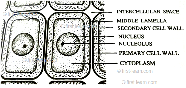
Cell Wall | Definition of Cell Wall |Middle Lamella|Primary, Secondary
Definition of cell wall- The thick and rigid , nonliving covering present just outside the plasma membrane of plant cells is called cell wall. It is discovered by Robert Hooke in 1665 while observing cell in the section of cork.
Explain about Cell Membrane | Structure of the Cell Membrane
Definition - Cell membrane is the thin, elastic and semipermeable living membrane that surrounds the protoplasm of a cell is called cell membrane o plasma membrane or plasma lemma.


From Cytoskeletal Structure to HOME PAGE


Recent Articles
-
Plants Around Us | Big & Small Plants | Shrubs & Herbs | Water Plants
Feb 03, 26 02:01 AM
 We see different types of plants around us. Plants are living things. They breathe and grow. They also reproduce. Most of the plants grow on land. Some plants grow in water.
 -
Formed Elements of Blood | Erythrocytes | ESR |Leukocytes |Neutrophils
Jan 15, 26 01:25 AM
 Formed elements formed elements are constitute about 45 % of blood afeias haematocrit value packed cell volume mostly of red blood corpuscles and are of 3 types- erythrocytes, leukocytes and blood pla…
 -
What Is Plasma? | Blood Plasma | Proteins | Nutrients | Cholesterol
Nov 07, 25 10:29 AM
 Blood is a mobile fluid which is a connective tissue and is derived from the mesoderm like cell any other connective tissue. Colour of blood is reddish and that flows inside the blood vessels by means…
 -
Disorders of Respiratory System | Tuberculosis | Pleurisy | Emphysema
Oct 28, 25 11:39 PM
Tuberculosis is very common disease and is caused by a type of bacteria called Mycobacterium tuberculosis. This disease causes different trouble in the respiration and infection of several parts of th… -
Regulation of Respiration | Respiratory Centres | Inspiratory Area |
Oct 14, 25 12:13 AM
 Respiratory Centre is the area that controls the rate of respiration and it is observed to be located in medulla oblongata and pons. Respiratory Centre has the following will dispersed components like…
New! Comments
Have your say about what you just read! Leave me a comment in the box below.